Xeroderma Pigmentosum Pdf
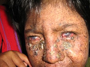
Xeroderma pigmentosum (XP) is an inherited condition characterized by an extreme sensitivity to ultraviolet (UV) rays from sunlight. This condition mostly affects the eyes and areas of skin exposed to the sun. Some affected individuals also have problems involving the nervous system. Symptoms typically develop by the time a child is 2 years old. Xeroderma pigmentosum is caused by mutations in genes that are involved in repairing damaged DNA. Inherited mutations in at least nine genes have been identified. The condition is inherited in an autosomal recessive manner.
People with XP need total protection from sunlight. This includes protective clothing, sunscreen, and dark sunglasses when out in the sun. To prevent skin cancer, medications like retinoid creams may be prescribed. Skin cancers that do develop should be treated using standard practices. There is no cure for XP, but there are ways to prevent and treat some of the problems associated with it.
Some of the strategies employed in the management of XP include:. Protection from ultraviolet light. Frequent skin and eye examinations. Prompt removal of cancerous tissue. Neurological examination. Psychosocial careSmall, premalignant skin lesions such as can be frozen with.
Larger areas of sun-damaged skin can be treated with or. In rare cases, therapeutic is used to remove damaged superficial epidermal layers. Skin cancers can be treated using standard treatment protocols, including electrodesiccation and curettage (scrapes away the lesion and uses electricity to kill any remaining cells), surgical excision, or chemosurgery. High dose oral or acitretin can be used to prevent new cancers. Cancers of the eyelids, and cornea are usually treated surgically. Corneal transplantation may be necessary for those with severe keratitis and corneal opacity.More detailed information about the treatment of XP may be accessed through the following online resources:. GeneReviews:. Medscape Reference - &.
Unusual changes in the melanin pigmentary system were observed on a warty papule biopsy taken from a patient with xeroderma pigmentosum (XP). Degenerated melanocytes full of lipids were observed from the basal layer up to the middle of the epidermis. The melanosomes were polymorphous, variable in size and shape with very strange aspects, such as spider-like and whirling configurations. The structure of the macromelanosomes suggests that they are large autophagosomes. These findings are discussed and compared with previous studies.

1.Califano A, Caputo R (1968) La microscopia elettronica nello studio dells precancerosi epiteliali cutanee. Minerva Dermatol 43:568–616. 2.Caputo R, Califano A (1971) Ultrastructural changes in the epidermis of xeroderma pigmentosum lesions in various stages of development. Arch Dermatol Forsch 241:364–373. 3.Cesarini JP, Bioulac P, Moreno G, Prunieras M (1975) Hypopigmented macules of sun unexposed skin in xeroderma pigmentosum.

An electron microscopic study. J Cut Pathol 2:128–139. 4.Clark WH, Bretton R (1971) A comparative fine structural study of melanogenesis in normal human epidermal melanocytes and in certain human malignant melanoma cells. In: Helwig EB, Mostoki FK (ed) The skin.
Williams Wieking Company, Baltimore, pp 197–214. 5.Cleaver JE (1968) Defective repair replication of DNA in xeroderma pigmentosum. Nature 78:652–656. 6.Cleaver JE (1972) Xeroderma pigmentosum: Variants with normal DNA repair and normal sensitivity to ultraviolet light.
J Invest Dermatol 58:124–128. 7.Guerrier CJ, Lutzner MA, Devico V, Prunieras M (1973) An electron microscopical study of the skin in 18 cases of xeroderma pigmentosum.
Dermatologica 146:211–221. 8.Hashimoto K (1975) A case of halo nevus with effete melanocytes. Acta Derm Venereol 55:87–95. 9.Hirone T, Eryu Y (1978) Ultrastructure of giant pigment granules in lentigo simplex. Acta Derm Venereol 58:223–229.
10.Hunter JAA, Zaynoun S, Paterson WD, Bleehen SS, Mackie R, Cochran AJ (1978) Cellular fine structure in the invasive nodules of different histogenetic types of malignant melanoma. Br J Dermatol 98:255–272. 11.Jimbow K, Szabo G, Fitzpatrick TB (1973) Ultrastructure of giant pigment granules (macromelanosomes) in the cutaneous pigmented macules of neurofibromatosis. J Invest Dermatol 61:300–309. 12.Jung EG (1978) Xeroderma pigmentosum; heterogeneous syndrome and model for UV carcinogenesis. Bull Cancer 65:315–321. 13.Konrad K, Wolff K, Honigsmann H (1974) The giant melanosome: A model of deranged melanosome-morphogenesis.
J Ultrastruct Res 48:102–123. 14.Lerner AB (1971) On the etiology of vitiligo and gray hair. Am J Med 51:141–147. 15.Morohashi M, Hashimoto K, Goodman TF, Newton DE, Rist T (1977) Ultrastructured studies of vitiligo, Vogt-Koyanagi syndrome, and incontinentia pigmenti achromians. Arch Dermatol 113:755–766. 16.Ortonne JP, Perrot H, Schmitt D, Bioulac P (1980) (in press) Cutaneous hypermelanosis and intramelanocytic lipid droplets.
Arch Dermatol. 17.Rasheed A, El Hefnawi H, Nagy G, Wiskemann A (1969) Elektronenmikroskopische Untersuchungen bei Xeroderma pigmentosum. Arch Klin Exp Dermatol 334:321–344. 18.Takahashi M, Fitzpatrick TB (1977) Formation of macromelanosomes in melanocytic naevi.

Xeroderma Pigmentosum Pdf Cover
Presented at the Xth International Pigment Cell Conference. Cambridge, Massachusetts. 19.Tsuji T (1974) Electron-microscopis studies of xeroderma pigmentosum: Unusual changes in the keratinocyte. Br J Dermatol 91:657–666.